
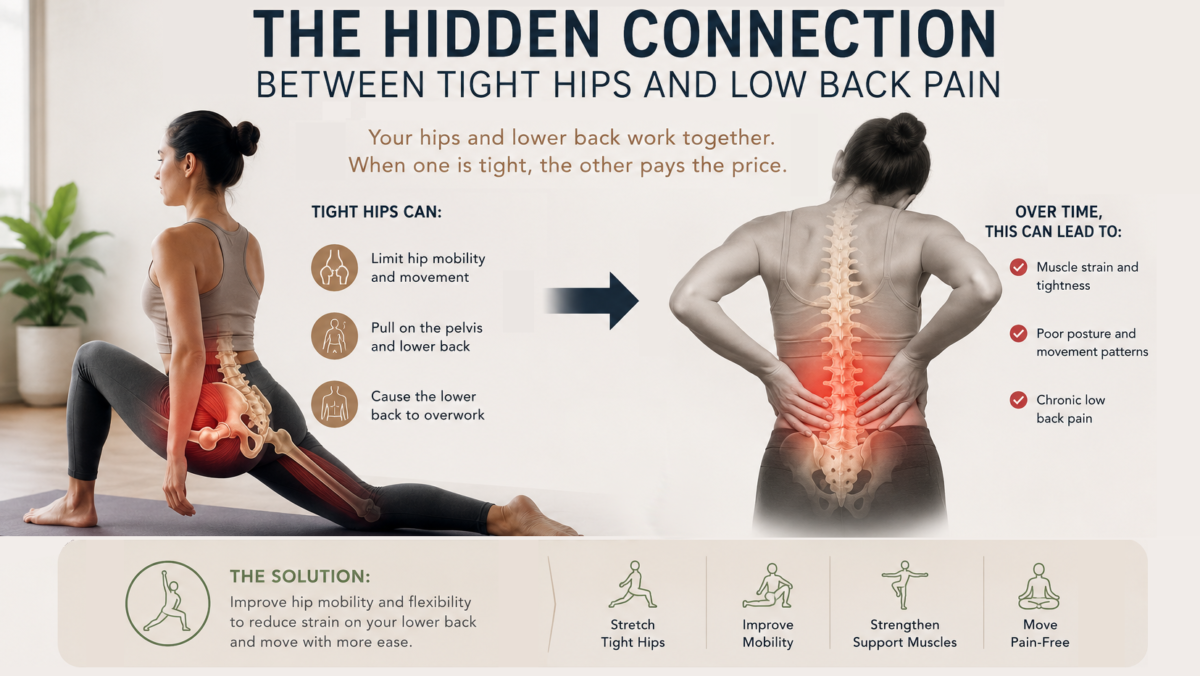
Why Your Hips May Be the True Catalyst for Lower Back Pain
If you have been searching for solutions to chronic lower back pain, you might find it counterintuitive that the primary driver of your discomfort may not reside in your spine at all. Instead, a pervasive cause of persistent lumbar distress is a biomechanical restriction located just below it: restricted hip mobility.
In clinical soft-tissue therapy, a recurring physiological pattern manifests across diverse populations, from elite collegiate athletes to sedentary office workers. Patients frequently present with primary complaints of lower back pain, yet diagnostic movement assessments routinely trace the culprit back to structural restrictions and muscular imbalances within the hip complex. Understanding this lumbopelvic connection is essential for establishing long-term therapeutic relief.
The Biomechanics of Lumbopelvic Compensation
The hip is a ball-and-socket joint engineered to be one of the most mobile structures in the human body, facilitating multi-planar movements such as flexion, extension, abduction, adduction, and rotation.
According to the regional interdependence model of musculoskeletal rehabilitation, dysfunction in one anatomical region can induce compensatory stress in an adjacent segment (Roach et al., 2015). Range of motion (ROM) is severely compromised when the soft tissue surrounding the hip joints becomes pathologically tight. To maintain a functional gait and upright posture, the kinetic chain must compensate. It achieves this by forcing the lumbar spine—a region evolutionarily designed for stability rather than extreme mobility—to hyper-extend and rotate excessively.
Over time, this secondary mechanical compensation places microtraumatic stress on the lumbar vertebrae, deep spinal musculature, and intervertebral discs. This biomechanical overload manifests clinically as:
- Myofascial chronic muscle tightness and acute spasms
- Facet joint irritation due to excessive compressive forces
- Compromised core stability and altered postural alignment
- Altered neural dynamics mimicking sciatica
The Hip Flexors: Structural Adaptation to Sedentary Lifestyles
The primary muscular driver of this dysfunction is typically muscle tightness in the hip flexor complex, primarily the iliopsoas and rectus femoris. The primary anatomical function of these muscles is to decrease the angle between the leg and the hip.
Modern daily routines create an environment highly conducive to hip flexor shortening. Prolonged sitting—whether studying at a desk, driving, or engaging in sedentary screen time—keeps these muscles in a chronically shortened, non-functional state. Over time, the tissue undergoes adaptive shortening, altering its resting length.
When an individual attempts to stand or walk with shortened hip flexors, these muscles pull you down into a bent over position. This mechanical pull can induce an anterior pelvic tilt (APT), which artificially exaggerates lumbar lordosis (the inward curvature of the lower back) and increases shear stress on the lower lumbar segments (Laird et al., 2014).
Secondary Musculotendinous and Fascial Contributors
While the hip flexors serve as a primary catalyst, the lumbopelvic-hip complex (LPHC) involves several other interconnected soft-tissue structures:
- The Gluteal Complex: The gluteus maximus and medius are critical for hip extension and lateral pelvic stabilization. When hip flexors are chronically tight, they neurologically suppress the glutes through a mechanism known as reciprocal inhibition. Weak or deactivated glutes force the hamstrings and lower back muscles to take over during hip extension, leading to early muscle fatigue and strain (Bade et al., 2014).
- The Piriformis: This deep lateral rotator runs in close anatomical proximity to the sciatic nerve. Tightness of the piriformis can entrap or compress the nerve, inducing piriformis syndrome, which presents as radiating pain, numbness, or tingling down the posterior leg (Hopayian et al., 2018).
- The Fascial Network: Muscles do not operate in isolation; they are enveloped in fascia, a continuous web of dense connective tissue. Chronic tension or inflammation creates fascial adhesions that restrict multi-directional tissue glide, binding down muscles and propagating structural tension patterns throughout the entire lower kinetic chain.
Limitations of Conventional Static Stretching
When experiencing tightness, the standard response is often prolonged static stretching. However, clinical data indicates that stretching a chronically tight muscle in isolation rarely yields permanent structural changes if underlying neuromuscular patterns are ignored.
Tightness is frequently a protective neurological response rather than a literal shortening of muscle fibers. If a joint lacks stability or exhibits muscle weakness elsewhere, the nervous system will actively signal surrounding muscles to tighten to prevent joint damage. Consequently, simply stretching the tissue without addressing structural imbalances, fascial restrictions, or poor movement mechanics provides only transient symptom management.
Targeted Manual Therapies and Neuromuscular Re-education
To achieve definitive resolution, therapeutic interventions must address both mechanical tissue restrictions and neurological patterning:
- Myofascial Release & Deep Tissue Assessment: Targeted manual therapy breaks up fascial adhesions and triggers localized vasodilation (increased blood flow), restoring the tissue’s natural elasticity and fluid dynamics.
- Active Isolated Stretching (AIS): Unlike traditional static stretching, AIS utilizes short, dynamic, controlled contractions of the opposing muscle group held for no more than two seconds (Medeiros & Martini, 2018). This specialized modality capitalizes on the body’s natural neurophysiology: contracting the agonist muscle automatically signals the target antagonist muscle to relax, allowing for safe, incremental increases in range of motion without triggering the protective muscle spindle stretch reflex.
Conclusion: Shifting the Clinical Paradigm
The clinical relationship between restricted hip mechanics and lower back pathology highlights a fundamental rule of human movement: the site of chronic pain is rarely the source of the problem. For athletes seeking to optimize athletic performance, or students enduring extensive hours of lecture-hall sitting, resolving chronic lower back distress requires looking beyond the spine. By restoring normal joint movement, muscular balance, and tissue flexibility within the hips, you effectively remove the compensatory demands placed on the lower back, addressing the root cause of the dysfunction rather than merely managing its symptoms.
At Simo Massage, my goal is simple: Don’t just feel good. Feel better. Through therapeutic massage, sports massage, myofascial release, and Active Isolated Stretching, I help clients uncover the root cause of pain and get back to moving the way their bodies were designed to move.
References
- Bade, M., Cobo-Estevez, M., Neeley, D., Pandividi, J., Osterhues, J., & Cleland, J. A. (2014). Effects of manual therapy and exercise targeting the hips in patients with low back pain: A systematic review. Journal of Evaluation in Clinical Practice, 20(6), 851–857.
- Hopayian, K., Song, F., Riera, R., & Sambandan, S. (2018). The clinical features of piriformis syndrome: A systematic review. European Spine Journal, 27(12), 2933–2941.
- Laird, R. A., Gilbert, J., Kent, P., & Keating, J. L. (2014). Comparing lumbo-pelvic kinematics in people with and without non-specific chronic low back pain: A systematic review and meta-analysis. BMC Musculoskeletal Disorders, 15(1), 1–15.
- Medeiros, D. M., & Martini, T. F. (2018). Chronic effects of different types of stretching on ankle dorsiflexion range of motion: Systematic review and meta-analysis. Foot and Ankle Surgery, 24(3), 188–197.
- Roach, S. M., San Juan, J. G., Suprak, D. N., Lyda, M., & Bies, A. J. (2015). Passive hip range of motion is reduced in active subjects with chronic low back pain compared to controls. International Journal of Sports Physical Therapy, 10(1), 13–20.